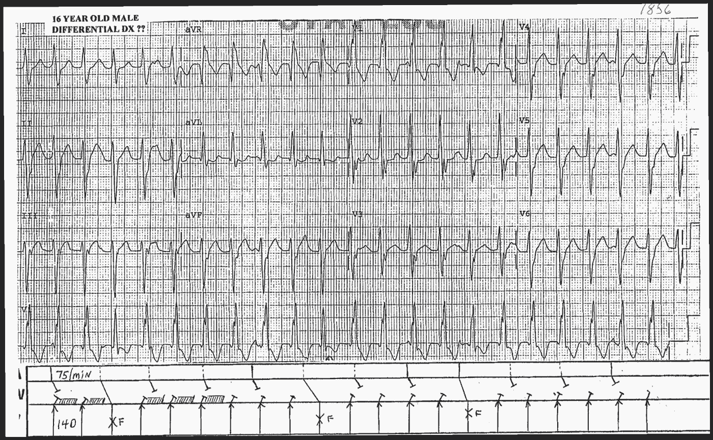
Interpretation
- Regulary Regular wide QRS complex Tachycardia
- RBBB Morphology of the QRS Complexes
- Left Axis Deviation (LAD)
- Some P waves are seen, so we have AV dissociation (seen in VT)
- QRS Complexes 4, 11 and 16 are with different morphologies because they are fusion complexes (seen in VT)
On first glance this tachycardia looks like supraventricular with abberancy (RBBB), but because of the AV dissociation and the fusion complexes, the diagnosis is Ventricular Tachycardia , most likely Idiopathic Posterior Fascicular Ventricular Tachycardia (VT).