This post is an answer to the Case – Generalized Bone Pain
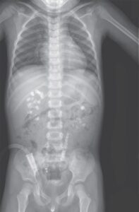
What abnormalities do you see on these selected radiographs ?
- Skull : Multiple well-defined lucent lesions affecting the cranial vault
- Ribs : Osteopenia and rib fractures
- Femur : Osteopenia, cortical thickening and focal cortical lucencies
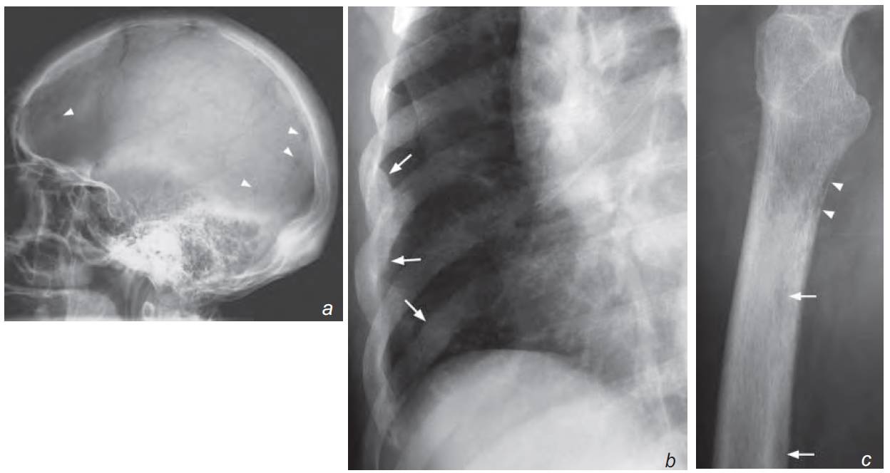
a) Lateral view of the skull shows multiple small punched-out (well-defi ned) lucent lesions (arrowheads)
b) Oblique view of right lower ribs shows generalized osteopenia and rib fractures (arrows)
c) Lateral view of the proximal right femur shows generalized osteopenia with tunneling (arrowheads). More discrete lesions are also seen (arrows)
Discussion
Multiple myeloma (diffuse malignant plasma cell proliferation) is the most common primary malignant bone tumour and occurs mostly in patients over 50 years of age. Bones containing red marrow are particularly vulnerable and are targeted in a radiographic ‘skeletal survey’.
- These bones include: flat bones (skull, pelvis, ribs etc.), lower thoracic lumbar spine and proximal femur and humerus. Radiographs show evidence of bone marrow infiltration such as:
- Multiple lucent lesions in bones
- Destruction of cortex
- Generalized osteopenia
Multiple myeloma and metastases are the two most common causes of multiple bone lesions in elderly patients. Multiple myeloma is a neoplastic process of plasma cells and involves bone marrow. However, in most cases, it does not elicit much osteoblastic response. Therefore a bone scan in these patients is usually normal (no hot spots) and thus it is not used for evaluating disease extent (as opposed to the evaluation of bone metastases).
This lack of osteoblastic response is less common with other neoplastic/metastatic diseases. As a haematological neoplasm, complications may arise due to thrombocytopenia (i.e. haemorrhage), hyper-coagulability (i.e. infarction) or leucopenia (i.e. infection).
Plasmacytoma is a focal form of malignant plasma cell proliferation. It tends to produce a large lesion affecting a single bone/area.
Role of imaging in patients with multiple myeloma
- Radiographs as a survey to monitor disease extent and complications (fracture)
- MRI and/or CT for complications (haemorrhagic, infections, etc.)
Key Points
- Multiple myeloma should be considered in elderly patients with multiple osteolytic lesions.
- Diagnosis is based on combination of laboratory and bone marrow biopsy results.
- Radiographic skeletal surveys help support the diagnosis and assess the extent of skeletal involvement.
- Generalized osteopenia and multiple osteolytic lesions are the typical radiologic features.