This post is an answer to the Case: 27-year-old woman with chest tightness and dyspnea
Findings
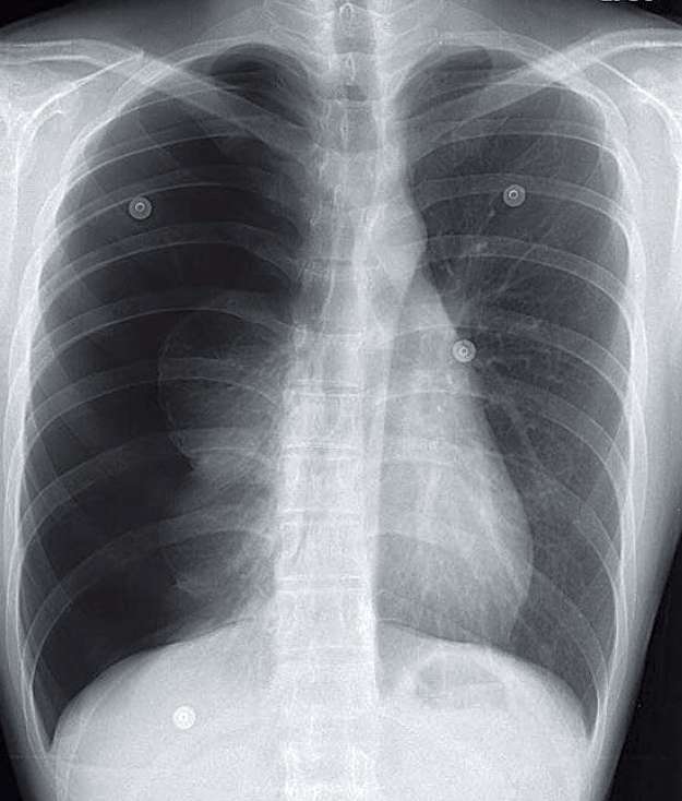
- Initial chest radiograph clearly demonstrates increased lucency in the right hemothorax with minimal displacement of the right hemidiaphragm inferiorly and shift of the mediastinum left ward. Note the increased distances between the ribs on the right, compared with those on the left. The right lung is partially collapsed.
- Visceral pleural surface can be seen as a thin white line, allowing distinction from a skin fold. No pulmonary vessels are seen lateral to the pleural line. The anterior junction line is also displaced left ward.
Differential Diagnosis
Tension pneumothorax. There should be no differential diagnosis.
Teaching Points
- A tension pneumothorax results in shift of the mediastinum to the contralateral side and the diaphragm inferiorly.
- In patients on positive ventilation, the diaphragm may be more displaced than the mediastinum, as the positive ventilation bolsters the unaffected side.
Management
- Needs emergent drainage. The mass effect can impede oxygen exchange on the contralateral side and impair venous return to the heart. Left untreated, a tension pneumothorax can result in cardiorespiratory collapse.
- When encountered in the reading room, the tension pneumothorax should prompt a phone call to the referring clinician. This phone call should be documented in the radiology report.
SIMILAR CASES:
Further Reading
- Barton ED. Tension pneumothorax. Curr Opin Pulm Med. 1999 Jul; 5 (4): 269 – 274. Leigh-Smith S, Harris T. Tension pneumothorax— time for a re-think? Emerg Med J. 2005 Jan; 22 (1): 8 – 16 .