This article is an answer to the Case – Infant with Vomiting and Abdominal Mass
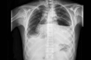
An upper gastrointestinal radiographic series was obtained (ultrasonography was not readily available as the initial test), and the findings showed a distended, air-filled stomach with undulating contours, known as the “caterpillar” sign.
This sign is visible when active hyperperistaltic waves come to an abrupt stop at the pylorus — a finding that is suggestive of hypertrophy.
Abdominal ultrasonography was then performed, and the results showed a pyloric wall width of 4.5 mm, a pyloric diameter of 25.0 mm, and a pyloric channel length of 24.0 mm.
Given that a pyloric wall width of 3 mm is considered to be the cutoff for the normal range, the 4.5-mm value in this infant confirmed the diagnosis of infantile hypertrophic pyloric stenosis.
The infant underwent an open pyloromyotomy and had no complications from the procedure. He was discharged home 2 days later and was able to receive full feedings. At follow-up 3 months later, he remained well.
References
- Frank Chen, Joseph Cernigliaro, Shweta Bhatt. (2021) The caterpillar sign. Abdominal Radiology 46:1, 394-395.
- Bshara Mansour, Adib Habib, Hussein Shamaly, Zahi Abou Nassar, Ahlam Abu Ahmad. (2020) Temporal caterpillar sign in a case of infantile hypertrophic pyloric stenosis. Pediatrics International 62:5, 654-655.
- Reiko Kawai, Kazuhiro Uda, Yuho Horikoshi, Hiroshi Hataya. (2019) Caterpillar Sign in an Infant with Hypertrophic Pyloric Stenosis. The Journal of Pediatrics 208, 292.
- Vikas Shah, Rohit Sharma. 2017. Caterpillar sign (pyloric stenosis). Radiopaedia.org.