This post is an answer to the Case – Unconscious After Head Injury
What does the CT show ?
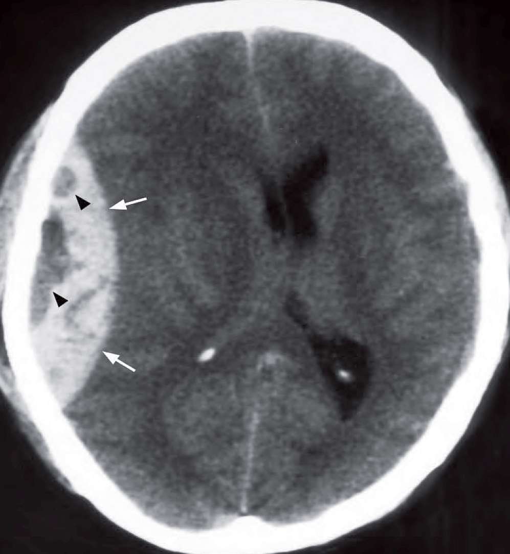
Extra-axial blood in the right temporoparietal area, heterogeneous in density, biconvex in shape with mass effect causing cerebral and ventricular compression and midline shift to the left.
What is the diagnosis ?
Non-contrast axial CT of the brain showing an acute right frontoparietal extradural haematoma (arrows) with mass effect on the underlying brain and lateral ventricles, midline shift to the left. The low density areas (arrowheads) within the extradural indicate active bleeding.
Discussion
The bleeding is often from a tear in the right middle meningeal artery and may be associated with a fracture.
Bleeding sources of extradural haematoma:
- Arterial (90%): tear of the middle meningeal or other meningeal arteries
- Venous (10%): sinus laceration, tear of meningeal vein
CT features of EDH:
- Biconvex/lenticular in shape
- The haematoma does not cross the coronal and lambdoid sutures
- May cross dural reflections falx cerebri and tentorium cerebelli
- Most are associated with skull vault fractures
- Low density areas within the EDH indicate active bleeding and thus heterogeneity may predict rapid expansion of the haematoma
- Venous EDHs from low pressure bleed are more variable in shape and may have delayed onset.
- The presence/absence of skull vault fracture does not predict the presence/ absence of intracranial injury.
- Skull radiographs have limited value in patients with severe trauma.
- In addition to the identifi cation of blood one must also evaluate the heterogeneity within the haematoma, extent of the mass effect, ventricular dilatation if any, the presence of fractures and evidence of any other intracranial injury.
SIMILAR CASES: