This is Answer to the Case Here .
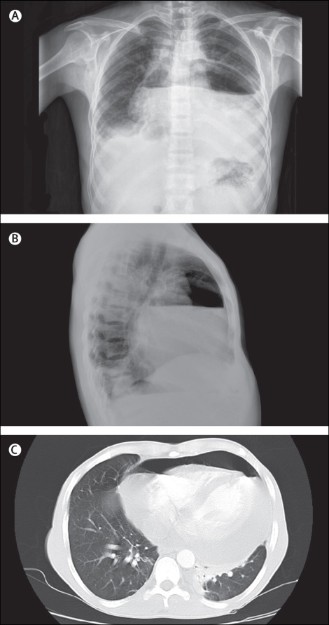
Chest radiography indicated cardiomegaly with an air–fluid level at the level of the heart.
Chest CT showed hydropneumopericardium. Echocardiography showed a massive pericardial effusion causing more than 25% respiratory variation of mitral inflow, consistent with tamponade physiology.
A pericardial window was made and drained about 1 L of serosangiunous fluid. Pericardial fluid culture was negative and pericardial biopsy analysis showed acute and chronic inflammation and fibrosis.
The patient had an uneventful hospital course after therapeutic drainage. 2 weeks later, follow-up echocardiography showed only mild pericardial effusion.