This article is an answer to the Case – Malaise, Back Pain, Dyspnoea and Cough with Purulent Sputu
Peripheral WCC was elevated at 13.0 x 10⁹ / L with a neutrophilia of 11.94 x 10⁹ / L. CRP increased at 246 mg/L. Arterial blood gas on room air; PaO2 9.52. Sputum and blood cultures were negative.
Imaging Findings
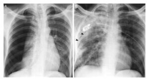
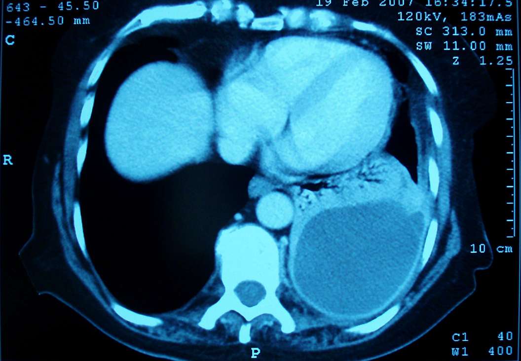
Chest X-ray (CXR) was reported as showing a cystic lesion at the left base with an air-fluid level seen within it. Contrast enhanced CT scan of chest demonstrated a fluid and air-filled cavity at the left lung base with an enhancing thick wall on coronal and axial images (see image). Patient was diagnosed with a left lower lobe lung abscess.
Treatment was commenced with intravenous clindamycin and ciprofloxacin. The patient improved dramatically within twenty four hours of commencing antibiotic therapy . CXR after two weeks antibiotic therapy demonstrated a marked improvement in the left lower zone changes with a normalization of the WCC and CRP.
The patient was subsequently discharged to complete a further four weeks oral antibiotic therapy. Follow-up CT performed at 3 months showed almost complete resolution of the abscess on the coronal and axial images.
SIMILAR CASE: Multiple Pulmonary Bacterial Abscesses