This article is an answer to the Case – Multinodular Tumor of Left Foot
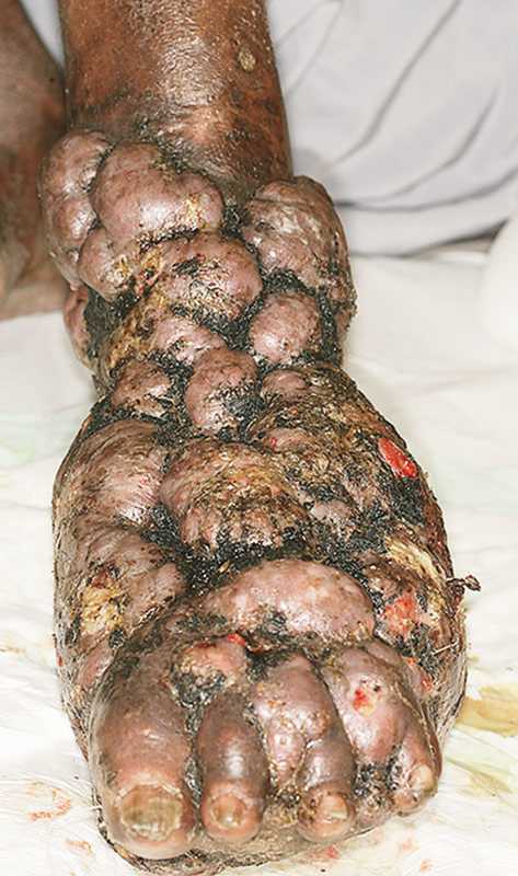
Examination revealed soft-tissue swelling; multiple painless, sometimes ulcerated, weeping tumefactions; and ipsilateral popliteal and inguinal lymphadenopathy. Magnetic resonance imaging revealed osteomyelitis of the tarsal bones.
The suspected diagnosis was a mycetoma, although Kaposi’s sarcoma and epithelioma cuniculatum (verrucous carcinoma) were also considered.
Pathological analysis of a punch-biopsy specimen with Giemsa staining revealed an inflammatory infiltrate surrounding granules with peripheral “clubs,” identified as actinobacteria. Culture of the specimens did not grow any organism.
Amikacin (for 10 days) and trimethoprim–sulfamethoxazole were given. After 1 month, the patient was discharged home and was able to walk with crutches. Ten months later, he was able to walk without assistance and had regained plantar sensitivity. Three years after beginning treatment, he was able to bend the toes and ankle.
Therapy with twice-daily trimethoprim–sulfamethoxazole (160 mg and 800 mg, respectively) has been maintained at the same dose since the beginning of treatment, and improvement of the lesions is still ongoing after 5 years of antibiotic therapy.