This post is an answer to the Case – A 77-year-old Woman with Progressive Memory Loss
What is shown on the CT ?
- Multiple, round, enhancing lesions.
- Perilesional oedema.
- Mass effect with compression of the left lateral ventricle and displacement of the midline structures to the right.
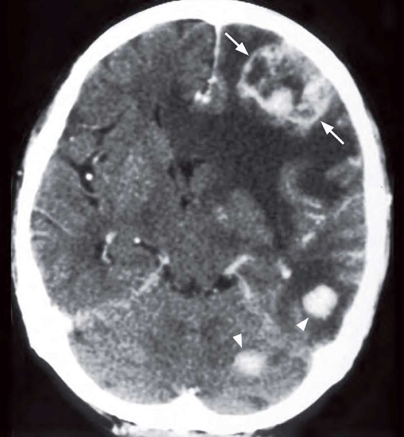
Diagnosis: Brain metastases
Axial contrast enhanced CT showing multiple enhancing metastases with perilesional oedema. Note irregular rim enhancement in the larger left frontal lobe metastasis.
Discussion
The most common primary tumours giving rise to metastatic brain deposits include:
- Bronchogenic carcinoma
- Breast carcinoma
- Colorectal carcinoma
- Renal cell carcinoma
- Melanoma
- Choriocarcinoma
- Leukaemia/lymphoma
- Neuroblastoma (in children)
Brain metastases are the result of haematogenous spread, are more likely to be multiple than solitary, and are of varying sizes.
The corticomedullary junction is the typical location and most are deposited in the cerebral hemispheres, followed by the cerebellum and brainstem
Usually associated with perilesional oedema that may be disproportionately large compared to size of the metastases.
In a patient with a known cancer the presence of white matter oedema on a noncontrast CT must be viewed with suspicion for metastatic disease (even if no obvious masses/nodules are seen). A post-contrast CT/MRI will improve the identifi cation of small metastases associated with the white matter oedema. However, in patients on steroid therapy the perilesional oedema may be missing.
Imaging findings on CT/MRI:
- Metastases appear as round intra-axial lesions of decreased attenuation on plain CT (except for haemorrhagic or calcified brain metastases) and high T2W signal on MRI
- Some primary tumours produce haemorrhagic metastases in the brain, and these include:
- Melanoma
- Choriocarcinoma
- Oat cell carcinoma of lung
- Renal cell carcinoma
- Thyroid carcinoma