This post is an answer to the Case – Dyspnea, Decreased Appetite, Fatigue and Fever
A chest radiograph showed a complete opacification of the left hemithorax with a deviation of the trachea and the heart to the right.
Ultrasound of the left hemithorax showed a minimal loculated pleural effusion and an extensive heterogeneous mass within the left chest cavity.
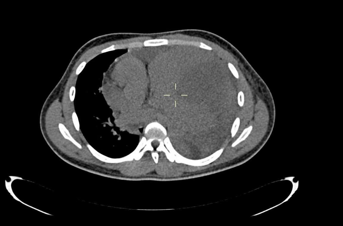
Thoracic and abdominal computed tomography with and without IV contrast demonstrated a 16 x 13 x 10 cm heterogeneous enhancing anterior mediastinal mass lesion without fat or calcifications but areas of necrosis. The mediastinal structures and heart were displaced towards the right side. There was a compression atelectasis of the left lung, a minimal pleural effusion on the left side and a moderate pericardial effusion.

CT guided needle biopsy of the mass revealed a large B-cell lymphoma.
Differential Diagnosis
- Primary mediastinal large B-cell lymphoma
- Thymoma
- Germ cell tumour teratoma
- Mediastinal thyroid
- Mediastinal tuberculosis
Discussion
For anterior mediastinal masses, the classic differential diagnosis is the “4 Ts”; namely thymoma, thyroid, teratoma and terrible lymphoma.
Germ cell tumours are common in young adults and constitute 15% of all anterior mediastinal masses in adults. Histologically they are divided into benign teratomas, seminomas and embryonal tumours which are also known as malignant teratoma or non-seminomatous tumour [5].
75% of all mediastinal germ cell tumours are mature teratomas. Calcification, teeth, bone and/or fat in a lesion are indicative of a teratoma. Serum tumour markers, AFP, beta HCG are also helpful for the diagnosis of a germ cell tumour [4, 5]
Mediastinal lymphomas are common, either as part of a disseminated disease or less commonly as the site of primary origin. The mediastinum is commonly affected by systemic lymphomas. Only 10% of lymphomas which involve the mediastinum are primary and the majority are Hodgkin lymphomas, accounting for 50-70%, while non-Hodgkin lymphoma make up 15-25% [3].
Primary mediastinal lymphomas are most frequently of three histologic varieties:
- Nodular sclerosing Hodgkin lymphoma
- Primary mediastinal large B-cell lymphoma
- Lymphoblastic lymphoma
Primary mediastinal large B-cell lymphoma is a sub-type of diffuse large B-cell lymphoma derived from the thymus [1, 3]. It represents less than 3 % of all non-Hodgkin lymphomas [2]. It is an aggressive neoplasm affecting a younger age group with female predilection.
CT demonstrates a smooth, lobulated soft tissue attenuating bulky mass, with areas of necrosis. Other features include parenchymal invasion, pleural effusion, pericardial effusion, chest wall invasion, tracheal, oesophageal and vascular compression. However, it is difficult to differentiate primary mediastinal large B-cell lymphoma from Hodgkin lymphoma and lyphoblastic lymphoma on the basis of imaging findings alone [1]. Histopathology and immunohistochemistry are necessary to distinguish these entities [2].
In our case the age of the patient, clinical features, radiological appearance, laboratory findings and histopathology along with immunohistochemistry helped to establish the final diagnosis of an anterior mediastinal large B cell lymphoma.
References
- K Shaffer, D Smith, D Kirn, W Kaplan, G Canellos, P Mauch and L N Shulman (1996) Primary mediastinal large-B-cell lymphoma: radiologic findings at presentation. AJR 167; 425-430 (PMID: 8686620)
- Johnson PW1, Davies AJ (2008) Primary mediastinal B-cell lymphoma. American society of hematology 2008.no 1;349-358
- Duwe BV1, Sterman DH, Musani AI (2005) Tumors of the mediastinum. chest 128(4):2893-2909 (PMID: 16236967)
- Brown LR1, Aughenbaugh GL (1991) Masses of the anterior mediastinum: CT and MR imaging. AJR Am J Roentgenol 157(6):1171-80. (PMID: 1950860)
- Kumar N, Gera C, Philip N. (2013) Isolated mediastinal tuberculosis: a rare entity. J Assoc Physicians India 61(3):202-3 (PMID: 24475683)


