This post is an answer to the Case – Long-Standing, Painful Deformation of the Right Tibia
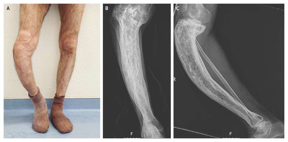
Physical examination revealed a prominent saber-like tibia and superficial venous circulation related to chronic venous insufficiency (image below, Panel A). Radiographs showed cortical thickening, trabecular enlargement, and bowing of the tibia (Panel B shows the anteroposterior view and Panel C the lateral view).
Levels of bone turnover markers were increased, including an alkaline phosphatase level of 345 U per liter (normal range, 45 to 129), an N-terminal propeptide of type I procollagen (P1NP) level of 506.3 ng per milliliter (normal value, <36.4), and a level of the β-isomer of the C-terminal telopeptide of type I collagen (β-CTX) of 1.38 ng per milliliter (normal value, <0.30). These findings show the clinical sequelae of prolonged early-onset Paget’s disease of bone.
Zoledronate at a single dose of 5 mg was prescribed owing to the increased metabolic activity in a weight-bearing location. Near-normalization of the alkaline phosphatase level (69 U per liter), the P1NP level (40.6 ng per milliliter), and the β-CTX level (0.34 ng per milliliter) was seen after 1 year, with symptomatic improvement.