This post is an answer to the Case – Recurrent Superficial Thrombophlebitis, DVT, and Non-Healing Ulcer
A 26-year-old woman presented with recurrent superficial thrombophlebitis on all four limbs, deep vein thrombosis involving the lower limbs, fever, a large fungating non-healing ulcer on the left cubital fossa (figure A), episodic abdominal pain, and weight loss for the past 4 months. The patient did not complain of oral or genital ulcers, ocular involvement, or altered bowel habits. Her blood pressure was 110/80 mm Hg; and her left radial pulse and left brachial pulse in the dorsalis pedis were absent, with carotid bruits heard on both sides.
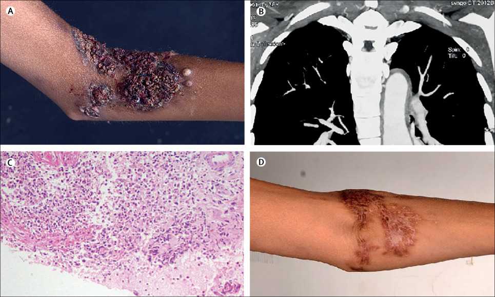
(B) Thickening and segmental dilatation of ascending aorta, arch, and descending thoracic aorta on CT angiogram.
(C) Dense acute necrotising suppurative inflammation comprised of neutrophil and nuclear debris on skin biopsy (haematoxylin and eosin stain, 20X).
(D) Healed ulcer with residual scarring after treatment.
The patient tested negative for antinuclear antibodies, antineutrophil cytoplasmic antibodies, antiphospholipid antibodies, and inherited thrombophilia. Pathergy and Mantoux tests were negative. Erythrocyte sedimentation rate was 60 mm/h and C-reactive protein concentration was 91 mg/L. Microbial cultures from the ulcer and blood were negative.
CT showed thickening and segmental dilatation of the ascending aorta, aortic arch, descending thoracic and abdominal aorta, and its major branches (figure B). Whole body 18F-fluorodeoxyglucose (18F-FDG) PET showed 18F-FDG avid circumferential wall thickening of the whole aorta and its major branches, without any organomegaly, lymphadenopathy, or lung involvement, suggesting active type V Takayasu arteritis.
Skin biopsy revealed suppurative necrotising (predominantly neutrophilic) inflammation of the deep dermis (figure C), mixed inflammatory infiltrates, secondary vasculitis (lymphocytic), absence of granulomas, and testing for acid-fast bacilli was negative, consistent with a diagnosis of pyoderma gangrenosum.
The patient was started on oral prednisolone (1 mg/kg) with monthly cyclophosphamide pulses (15 mg/kg) for 12 months. A marked improvement in symptoms and complete healing of the skin lesions with some scarring was seen after 6 months of therapy.