This post is an answer to the Case – Jaundice and Painless Neck Mass
Physical examination revealed a hard lymph node measuring 6 cm by 6 cm in the left supraclavicular fossa and hepatomegaly.
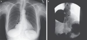
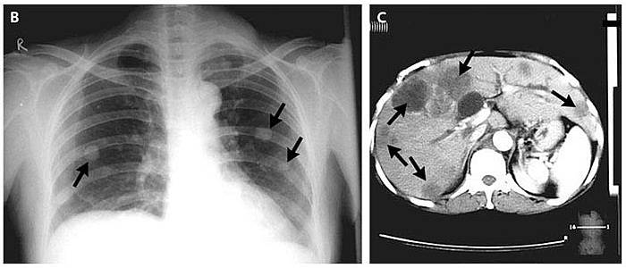
Plain radiography of the chest showed multiple nodular opacities in both lungs (Panel B, arrows). Computed tomography of the abdomen revealed multiple lesions in the liver (Panel C, arrows).
Endoscopy of the upper gastrointestinal tract showed a fungating mass around the ampulla of Vater. Biopsy specimens from the mass and the supraclavicular lymph nodes showed a periampullary adenocarcinoma with metastases.
The patient was treated with chemotherapy and palliative care. Abdominal cancers may metastasize to the left supraclavicular lymph nodes via the thoracic duct.