This post is answer to the ECG Case 240
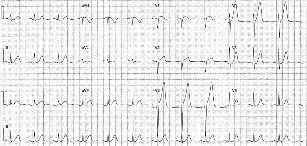
- Rate: 72 bpm
- Rhythm: Sinus rhythm
- Axis: Normal
- Intervals:
- PR – Normal (~160ms)
- QRS – Normal (80-100ms)
- QT – 320-360ms
- Segments:
- ST elevation in leads V1-4, maximal in V3
- ST elevation in lead III
- ST Depression in leads I, aVL, V5-6
- Additional:
- Hyperacute T-waves in leads II, III, aVF, V3-5
- Biphasic T wave V1
Interpretation
Antero-septal ST elevation with hyperacute T-waves. Likely LAD lesion, with possible ‘wrap-around’ component due to the inferior changes.
What happened next?
The patient was sent for urgent coronary angiogram which showed:
- 100% LAD occlusion → Stented
- 30% mid-RCA stenosis
We don’t have the full angio report so can’t comment on the exact location of the lesion or the anatomy of the LAD. The patient’s echo post procedure showed distal anteroseptal and anteroapical akinesis with preserved systolic function.
READ MORE: ECG Interpretation – All you need to know
SIMILAR CASES:


