This post is an answer to the ECG Case 334
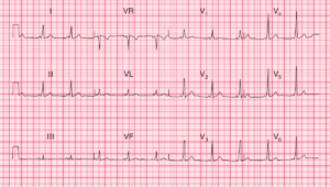
- Rate: 132 bpm
- Rhythm: Regular sinus rhythm
- Axis: Normal (-33 deg)
- Intervals:
- PR – Normal (~180ms)
- QRS – Normal (100ms)
- Additional:
- Voltage criteria LVH
- S V1 + R V5 = 35mm
- Non-voltage criteria LVH
- ST Depression in V5-6 with T wave inversion
- Features suggesting additional RVH
- Deep S wave in V5
- Biphasic high-voltage complexes in V4-5
- Features of left atrial abnormality
- Wide and deep P waves in lead V1
- Notching of P wave in lead II
- Features of right atrial abnormality
- Tall P wave in lead II
- Voltage criteria LVH
Interpretation
- Sinus tachycardia
- Features suggestive of four chamber enlargement
Note the ECG is poorly sensitive in detecting chamber enlargement and the terminology relating to p wave morphology now refers to atrial abnormality rather than atrial enlargement.
READ MORE: Left Ventricular Hypertrophy (LVH): How to Recognize it on ECG [With Examples]
SIMILAR CASES: