ECG Interpretation
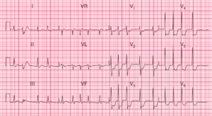
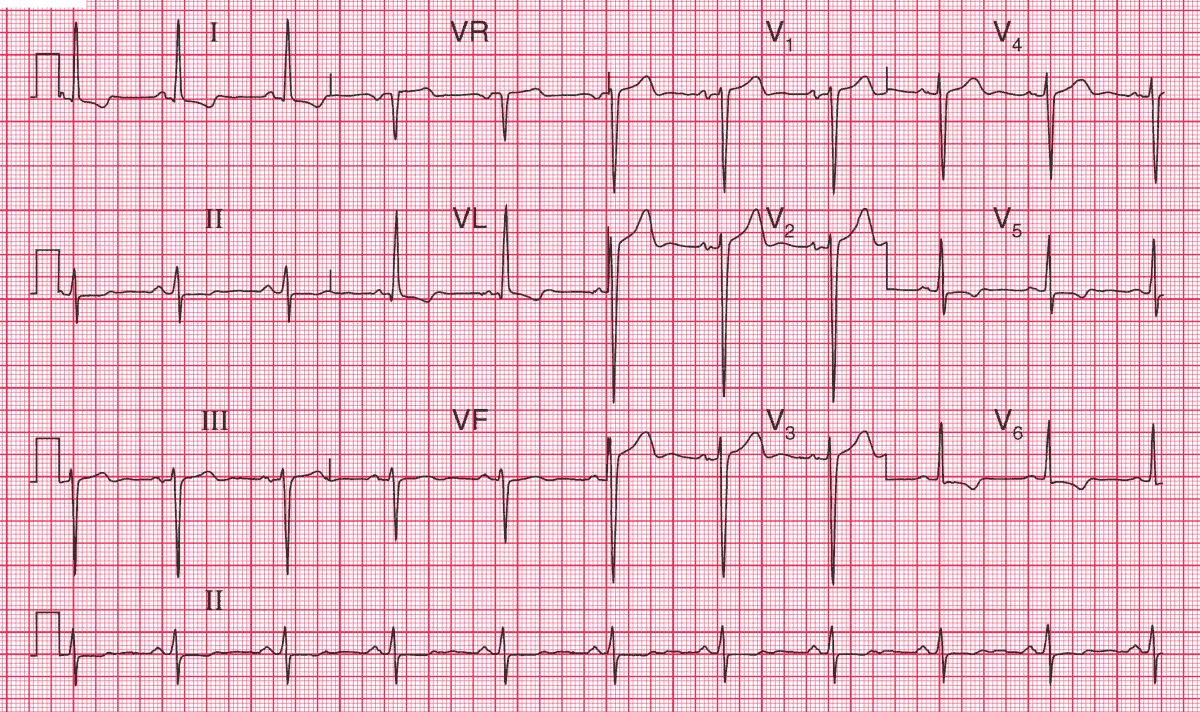
- Sinus rhythm, rate 60/min
- Bifid P wave (best seen in lead V3) suggesting left atrial hypertrophy
- Left ventricular hypertrophy by ‘voltage criteria’ (height of R wave in lead V6 plus depth of S wave in lead V2 = 50 mm)
- Lateral T wave inversion in lead I, aVL, V5 and V6 (left ventricular strain pattern)
Clinical interpretation
These are the classic changes of left atrial and left ventricular hypertrophy. In a patient with heart failure and a heart murmur the likely diagnosis is severe aortic valve disease.
What to do ?
The heart failure must be treated with diuretics, but it is essential to establish the cause of the left ventricular hypertrophy before selecting long-term treatment. It could be due to aortic stenosis or regurgitation, to mitral regurgitation or to hypertension.
While an angiotensin-converting enzyme inhibitor would be appropriate treatment for a patient with hypertension or mitral regurgitation, it would be potentially dangerous in a patient with aortic stenosis. An echocardiogram is the essential next step.
This patient had severe aortic stenosis, and needed an aortic valve replacement.
- READ MORE:
- Similar Cases: