This post is an answer to the Case – Skin Lesions with a Leopard-Like Appearance
Serum electrolytes were normal; C-reactive protein was raised to 40 mg/L (reference: <5 mg/L); and erythrocyte sedimentation rate was elevated at 46 mm/h (reference: <30 mm/h after 1 h), while complete blood count was within the normal range. Serum cortisol was 17 nmol/L (reference: 124–662 nmol/L) and adrenocorticotropic hormone (ACTH) was 274 pmol/L (reference: 2,6–10,1 pmol/L).
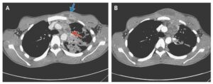
Based on her presentation, we suspected inadequate glucocorticoid replacement because of enhanced glucocorticoid demand. Hydrocortisone was increased to 40 mg per day. A CT scan showed splenomegaly and diffuse lymphadenopathy (figure B).
Surgical extirpation and histopathological analysis of an axillary lymph node revealed follicular lymphoma (stage 3B). The insidious development of lymphoma might have enhanced the patient’s glucocorticoid requirement, as reflected by her symptoms and elevated concentrations of ACTH.
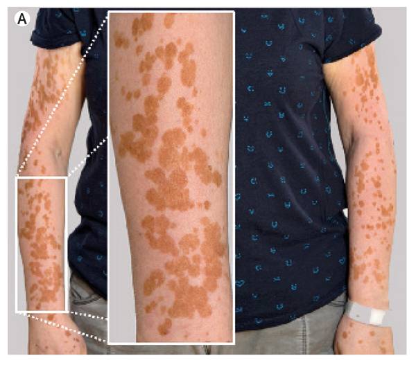
Cosecretion of α-melanocyte-stimulating hormone with ACTH, both from the common precursor POMC, enhanced the contrast of hyperpigmented islands on vitiligous skin.
After treatment with bendamustine and rituximab (R-Bendamustine protocol: 3 cycles, cumulative dose of bendamustine at 1080 mg [IV], cumulative dose of rituximab at 2250 mg [IV]), lymphadenopathy and splenomegaly had disappeared and the patient was free of symptoms when on hydrocortisone (20 mg per day).