The ECG shows a regular rhythm at a rate of 64 bpm. There is a P wave (+) before each QRS complex with a constant PR interval (0.20 sec). The P waves are positive in leads I, II, aVF, and V4-V6. This is, therefore, a normal sinus rhythm.
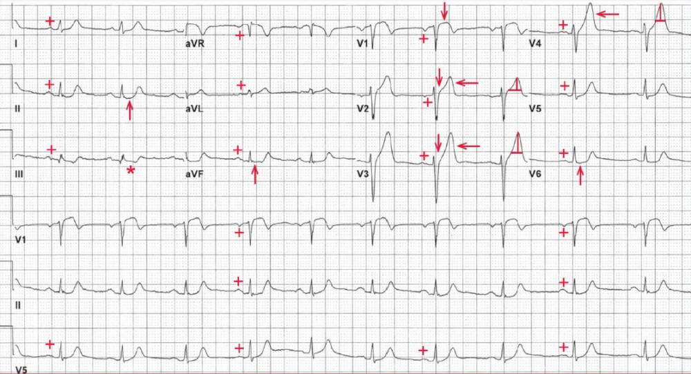
The QRS complex duration (0.08 sec) is normal and it has a normal morphology and axis, between 0° and +90° (positive QRS complex in leads I and aVF). The QT/QTc intervals are normal (380/390 msec).
Noted is ST-segment elevation in leads V1-V3 (↓) and slight ST-segment depression in leads II, III, aVF, and V6 (↑). In addition, the T waves in leads V1-V4 (<—) are tall and symmetric (hyperacute T waves).
This ECG is consistent with a very early acute anterior wall ST-segment elevation myocardial infarction (STEMI). The ST-segment depressions in leads II, III, aVF, and V6 are reciprocal changes.
Patients with STEMI require urgent revascularization, preferably with angioplasty and stenting. If not immediately available, a thrombolytic agent is indicated.
This patient did undergo cardiac catheterization, which revealed an acute thrombotic occlusion of the left anterior descending artery.
- READ MORE:
- SIMILAR CASES: