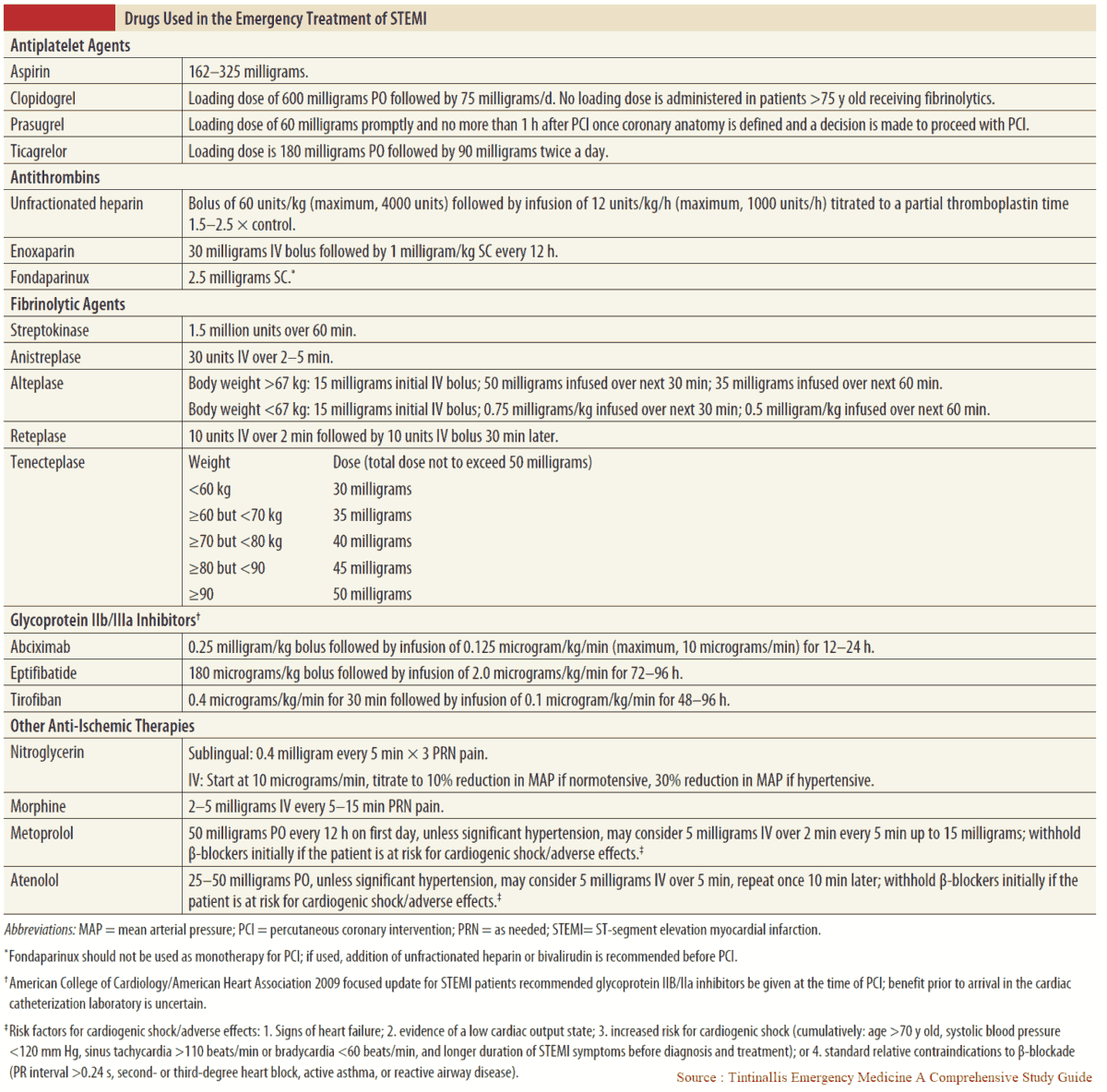
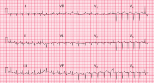
ECG Interpretation
- Sinus rhythm, rate 103/min
- Normal axis
- Q waves in leads II, III, VF
- Slow R wave progression in anterior leads
- Marked ST segment elevation in leads V1–V6 (Tombstone ST elevation)
- Hyperacute T waves in anterior leads

Clinical Interpretation
The Q waves in leads III and VF suggest an old inferior infarction, while the elevated ST segments, the hyperacute T waves and the slow R wave progression in leads V1–V6 indicate an acute anterior ST segment elevation infarction.
What to do next?
The patient should be given pain relief, and in the absence of the usual contraindications should immediately be treated with aspirin, immediate percutaneous coronary intervention (PCI) or a thrombolytic agent. If he was treated with streptokinase for his previous infarction, he should be given alteplase or reteplase on this occasion.
The drugs used in the treatment of Acute STEMI are summarized in the table below.