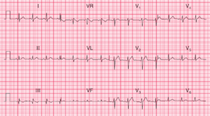
ECG Interpretation
- Sinus rhythm, rate 80/min
- One premature atrial contraction (PAC) – the sixth beat in lead II rhythm strip (circled)
- Left axis deviation (left anterior fascicular block)
- Poor R wave progression in precordial leads, with transition from S to R wave occuring in V5
- Normal QRS complexes, with a small Q wave (probably septal) in lead aVL
- Biphasic (up-down) T waves (terminal T wave inversion) in leads V1–V5 (spontaneous reperfusion)
Clinical Interpretation
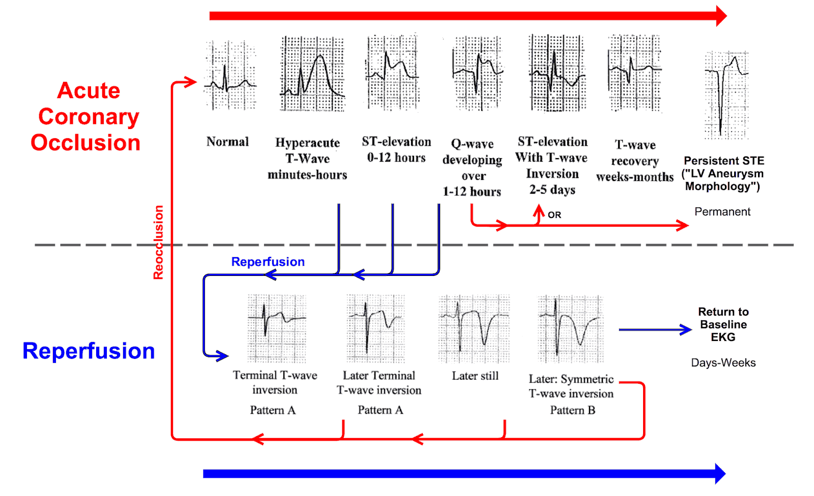
This is acute anterior MI with spontaneous reperfusion indicated by the biphasic T waves with terminal T wave inversion in leads V1–V5. In the image below you can see the evolution of MI with or without reperfusion.
What to do next?
The treatment is pain relief, aspirin, heparin, a beta-blocker and a statin – with PCI as soon as possible. The immediate outlook is good but the patient should be monitored and the ECG repeated to see if ST segment elevation is appearing or if the T waves become positive again which could indicated reocclusion.