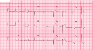
- Sinus Tachycardia
- Prominent P waves in inferior leads (P pulmonale) – a sign of Right Atrial Enlargement (RAE)
- Right Axis Deviation
- Flattened and Inverted P waves in lead I and aVL
- Poor R wave progression in precordial leads (delayed R/S transition)
- Low QRS voltage in lead I , aVL , V5 and V6
- One PVC
All of these ECG changes are seen in Chronic Obstructive Pulmonary Disease (COPD) .
Read More about :