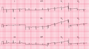
Right Axis Shift of P waves with prominent P waves (P Pulmonale – Right Atrial Enlargement (RAE)) in inferior leads (II , III , aVF) and flat or inverted P waves in lateral leads (I and aVL)
Right Axis Deviation
Low QRS Voltage in left precordial leads (V4-V6)
Poor R wave progression with delayed R/S transition in precordial leads, with sometimes persistance of S wave in V6 and absence of R waves in V1-V3 (“SV1-SV2-SV3” sign)
Right ventricular hypertrophy
Right bundle branch block
Multifocal atrial tachycardia
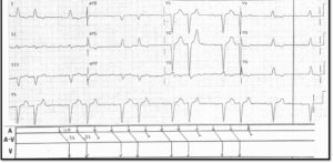
Example 1
ECG of Chronic Obstructive Pulmonary Disease (COPD)
Sinus Tachycardia
Prominent P waves in inferior leads (P pulmonale) – a sign of Right Atrial Enlargement (RAE)
Right Axis Deviation
Flattened and Inverted P waves in lead I and aVL
Poor R wave progression in precordial leads (delayed R/S transition)