ECG Interpretation
- Sinus rhythm, rate 72/min
- Normal axis
- Large Q waves in leads V1–V4 and small Q waves in leads I, VL
- Elevated ST segments and inverted T waves in leads V2–V5
- Flattened T waves in leads I and V6; inverted T waves in lead VL
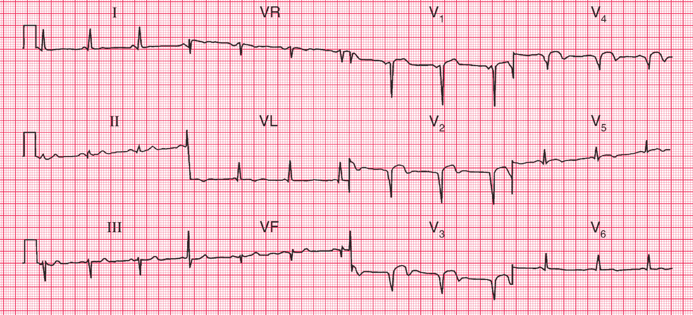
Clinical Interpretation
This ECG would be compatible with an acute anterior myocardial infarction, but this does not fit the clinical picture: it appears that an event occurred 2 months previously. This pattern of ST segment elevation in the anterior leads can persist following a large infarction, and is often seen in the presence of a ventricular aneurysm.
What to do?
An echocardiogram will show the extent of the aneurysm and whether the remaining left ventricular function is impaired, which it almost certainly will be. The patient should be treated with diuretics and an angiotensin-converting enzyme inhibitor, and surgical resection of the aneurysm might be considered.
- READ MORE: ECG Interpretation: All you need to know
- More Similar Cases: Old Anterolateral MI with Left Ventricular Aneurysm