This post is an answer to the ECG Case 245
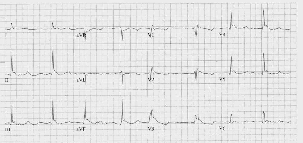
- Rate: ~48 bpm
- Rhythm:
- Irregular
- No p waves visible
- Axis: Normal
- Intervals:
- QRS – Prolonged (~180ms)
- QT – 720ms
- Segments: Inferior ST sagging
- Additional:
- RBBB Morphology
- Osborn J waves
- Prominent U waves best seen infero-laterally
- T wave inversion in leads aVR, aVL, V1-3
Interpretation
- Slow Atrial Fibrillation
- J-waves (Osborn waves)
- Prominent U waves
Differentials for this ECG
This ECG is most consistent with hypothermia but some features could be explained by drug toxicity (digoxin, CCB’s, beta-blockers), electrolyte abnormalities, ischemia, sinus node dysfunction.
We should be mindful in the elderly that the clinical situation is often multi-factorial and could be a combination of the above causes. Also remember hypothermia in the elderly has a multitude of potential causes including environmental, sepsis and endocrine.
READ MORE: Hypothermia Algorithm
SIMILAR CASES:


