This post is an answer to the ECG Case 296
- Rate: 72 bpm
- Rhythm:Regular sinus rhythm
- Axis: Normal (-33 deg)
- Intervals:
- PR – Short (80ms)
- QRS – Prolonged (120ms)
- QT – 400ms (QTc Bazette 380-400 ms)
- Segments: no significant changes
- Additional:
- Delta waves in leads I, aVL
- Deep Q wave in leads III, aVF
- T wave inversion in leads aVL, V1, V2, aVR
- Voltage criteria LVH
- R wave aVL > 11mm
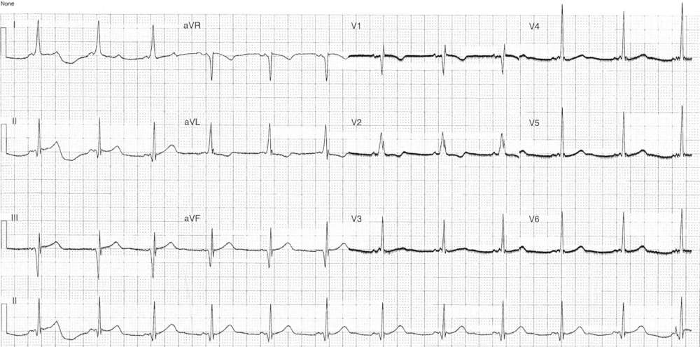
Interpretation
- Pre-excitation (WPW)
- Deep Q waves in inferior leads mimic old inferior MI referred to as pseudo-infarction pattern this is due to pre-excitation and does not reflect prior ischaemia.
- AP location is postero-septal tricuspid annulus using Arruda algorithm
What happened next ?
The patient had known WPW. She was investigated from a possible PE which was negative and discharged.
SIMILAR CASES: