This post is an answer to the ECG Case 319
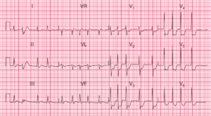
- Sinus rhythm, rate ~66 bpm
- Left axis deviation
- RBBB
- ST Elevation
- Lead III 1mm
- Lead aVF ~1mm
- Lead II – up-sloping ST
- ST Depression
- Leads V1-3, aVL
- Hyperacute T waves inferolateral leads
- Deep Q wave in leads III, aVF
Interpretation
Acute inferior OMI on a background of prior inferior MI
What happened next ?
The patient was taken for urgent angiography which showed:
- LMCA: Minor irregularities
- LAD: Long segment diffuse disease
- Cx: Patent stent, distal 70% stenosis
- RCA: Dominant vessel. Proximal occlusion of PLV branch → stented
The patient made an uneventful recovery and was discharge with out-patient cardiology follow-up.
SIMILAR CASE: ECG Case 60: Acute Anterior MI and Old Inferior MI
READ MORE: ECG Interpretation: All you need to know