Interpretation
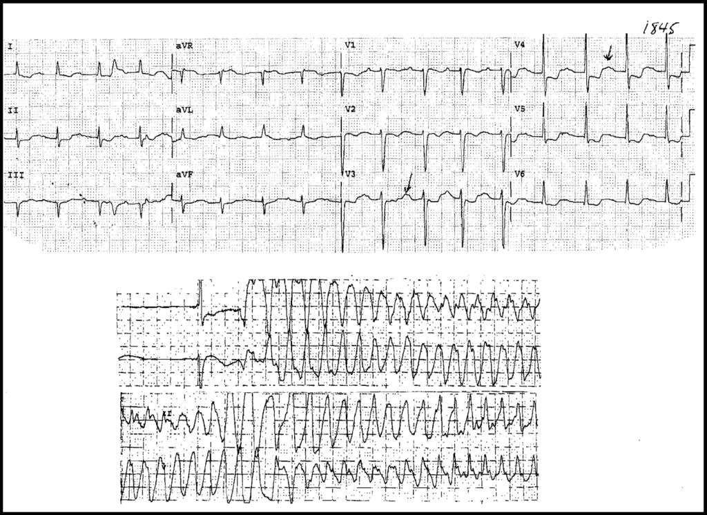
On the first ECG we see:
- Sinus rhythm approximately 100/min
- Prolonged PR Interval (First Degree AV Block)
- ST Depression in multiple leads
- U Waves best seen in precordial leads
- Prolonged QT (QU) Interval
- In leads I and III we see something that resembles a PVC, but because it is only present in these two leads it’s probably an artifact
All of these findings are pointing to Hypokalemia. Read more about Hypokalemia ECG Changes HERE.
On the second ECG we see Multiform Ventricular Tachycardia (Torsades de pointes) caused by the prolonged QT Interval from Hypokalemia.
In this Case the Hypokalemia was caused by excessive diuretic therapy.


