Hemoptysis – Causes, Evaluation and Management
Hemoptysis is the expectoration of blood from a subglottic source. Massive hemoptysis accounts for ~5% to 15% of cases and is often described as more than 600 mL of blood in a 24-hour period.
This 9-year-old boy presented with a two-day history of right shoulder pain after an upper respiratory tract infection. What is the cause of the abnormality demonstrated?
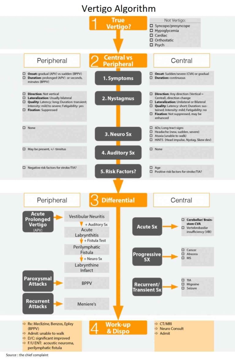
Emergency department evaluation relies on excluding life-threatening causes of Headache (killers), specifically SAH and meningitis. Then consider other serious causes of headache that can debilitate the patient (the maimers). When these have been excluded, we can then diagnose primary headache syndromes.
37-year-old man with Dyspnea 37-year-old man with known end-stage renal disease and chronic uraemic pericardial effusion presented with gradually worsening dyspnoea and orthopnoea, 4 days after a nephrectomy to control…
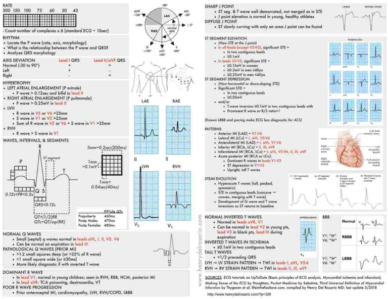
Right Axis Shift of P waves with prominent P waves (P Pulmonale - Right Atrial Enlargement (RAE)) in inferior leads (II , III , aVF) and flat or inverted P…
Ecg of a 81 Years old man. What disease can cause abnormalities like this on the ECG ?
Three hormones are available to the body for maintaining a calcium homeostasis: Vitamin D, Parathormone, Calcitonin
Sarcoidosis is a multisystem granulomatous disorder that is characterized by noncaseating granulomas. Pulmonary involvement is the most common manifestation, but extrapulmonary manifestations are seen in up to 30% of patients.
Work-up for upper abdominal pain begins with resuscitation and exclusion of critical diagnoses. Then, through a detailed history and physical, consider all the causes of upper abdominal pain. The differential may then come down to Gallstone etiology vs Gastritis/PUD → will need further imaging based on suspicion (US abdomen)
Most frequent ECG changes in Hypokalemia are: T waves flattening and inversion, U waves, long QT (QU) interval, ST depression and...