This post is an answer to the Case – Patient with Productive Cough for 1 Month
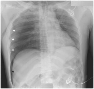
Chest X-Ray Interpretation
- Multiple areas of air-space opacification in the right lung also involving the apex
- Cavitation and air-fluid level within the opacified areas
- Blunting of right CP angle due to exudative effusion
Diagnosis: Caseous Necrosis in Tuberculosis
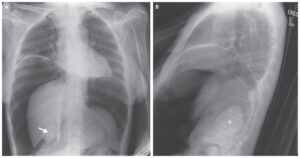
SIMILAR CASES: