ECG Intepretation
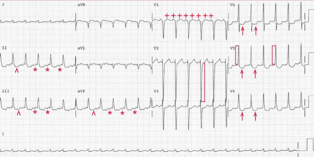
There is a regular rhythm at a rate of 150 bpm. Because the most common rate of atrial flutter is 300 bpm, atrial flutter with 2:1 AV conduction must be considered whenever there is regular supraventricular tachycardia at a rate of 150 bpm.
Distinct negative atrial waveforms can be seen in leads II, III, and aVF (*). Looking more closely at lead V1, two distinct positive atrial waveforms can be seen (+), one just before the QRS complex and the second superimposed on the T wave.
On closer inspection the second flutter wave can indeed be seen after the QRS complex in leads II, III, and aVF (˄), although initially it might have been considered an abnormal ST-T wave.
There is a regular PP interval, and the atrial rate is 300 bpm. Hence the rhythm is typical atrial flutter with 2:1 AV conduction.
The amplitude of the QRS complex is increased; the S-wave depth in lead V2 is 33 mm ( ] ) and the R-wave amplitude in lead V5 is 17 mm ( [ ) (S-wave depth in lead V2 + R-wave amplitude in lead V5 = 50 mm). The QRS complex amplitude is diagnostic for left ventricular hypertrophy.
There is also ST-segment depression in leads V3-V6 (↑), which is consistent with subendocardial ischemia that may be primary as a result of the rapid rate or secondary to left ventricular hypertrophy.


