This article is an answer and interpretation to the ECG Case 159
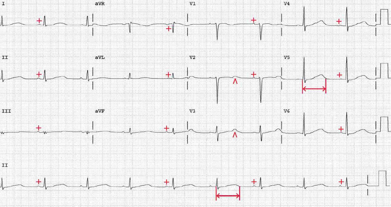
There is a regular rhythm at a rate of 52 bpm. There is a P wave (+) before each QRS complex with a stable PR interval (0.18 sec). The P wave is positive in leads I, II, aVF, and V4–V6 and negative in lead aVR. Hence this is a sinus rhythm.
Noted is a long QT interval (↔) (620 msec) and the rate-corrected QT (QTc) is 580 msec. As seen in leads V4–V6, there is a very broad T wave. Hence the QT prolongation is the result of prolonged depolarization, and this is a long QT syndrome (LQTS).
Also noted in leads V2 and V3 is a prominent U wave (^) that is on the T wave, interrupting it. This is called a QT-U wave and is seen in a congenital LQTS, particularly LQTS 1 and 2, which result from a channelopathy involving the potassium channel.
The QT interval is measured from onset of QRS complex (either a Q or R wave) to end of T wave. The lead used to measure the QT interval is the one in which the T wave is most distinct and where its termination is clear.
Identifying the termination of the T wave can be particularly difficult when a U wave is present. The U wave is not included in the QT measurement if it is after the T wave and distinct from and significantly smaller than the T wave. However, when the T and U waves are merged and the U wave is on top of the T wave or interrupts it, the U wave is included in the QT measurement.
The QT interval needs to be corrected for heart rate, using Bazett’s equation, QTc = QT interval ÷ square root of the RR interval (in sec) The normal corrected QT (QTc) ≤ 0.44–0.46 sec. It also should be remembered that the QT interval also includes the QRS complex.
The normal QT measurement is based upon a QRS complex duration that is normal (up to 0.10 sec). Therefore, if the QRS complex duration is increased, this needs to be considered in measuring the QT interval. The duration over 0.10 sec should be subtracted from the QT interval measurement and then the QT interval corrected for heart rate.
A long QT interval may be due to:
- Delayed repolarization in which the ST segment is long while the T wave is normal. This form of long QT interval is seen with metabolic abnormalities, particularly low calcium or magnesium.
- Prolonged repolarization in which the ST segment is normal but the T wave is broad. This may be due to drugs, termed an acquired QT prolongation, or a genetic abnormality producing a channelopathy, called congenital LQTS). It is congenital QT prolongation that may have a prominent U wave interrupting the QT interval (QT-U wave).
- READ MORE about Pearls in Syncope ECG Interpretation
- SIMILAR CASES:
- ECG Case 111: Marked Prolongation of the QT Interval – Long QT Syndrome
- ECG Case 104: Congenital Long QT Syndrome
- ECG Case 84: Drug-induced QT interval prolongation and polymorphic VT
- ECG Case 128: Torsade de Pointes (Nonsustained Polymorphic Ventricular Tachycardia Associated With a Long QT Interval)


