Hydropneumothorax as Initial Manifestation of Postprimary Tuberculosis

This post is answer to the Case – Patient with Weight Loss, Anorexia, Fever, Cough

Imaging Findings

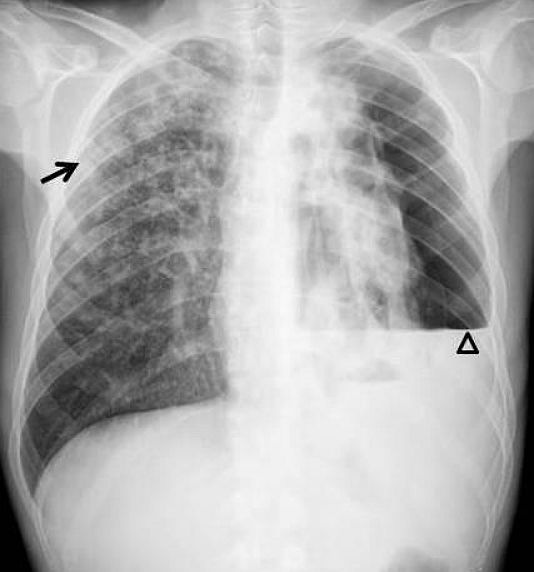
Posteroanterior radiography showed a hydropneumothorax on the left side with a collapsed left lung. On the contralateral side, ill-defined nodules and consolidations in the right upper lobe and upper segment of the lower lobe were seen (Fig. 1).

Chest radiograph reveals left-sided air-fluid level (arrowhead) with pneumothorax and consolidations in the right upper lobe and upper segment of the right lower lobe (arrow).
Fig. 1: Chest radiograph reveals left-sided air-fluid level (arrowhead) with pneumothorax and consolidations in the right upper lobe and upper segment of the right lower lobe (arrow).

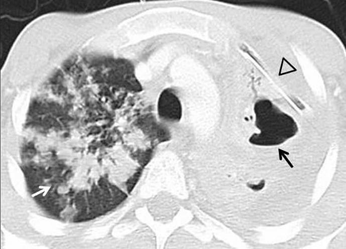
Tube thoracostomy was performed in the emergency department, with improvement of the symptoms. CT with intravenous contrast showed patchy areas of consolidation with air bronchogram, poorly defined margins, predominantly in the upper lobes. In addition, centrilobular nodules and the tree-in-bud pattern was observed. On the left side, several of these consolidations cavitated (Fig. 2).

CT Lung window setting reveals consolidations with cavities in the collapsed left lung (black arrow). The right upper lobe presents multiple consolidations (white arrow). Tube thoracostomy is positioned in the left pleural space(arrowhead).
Fig. 2: CT Lung window setting reveals consolidations with cavities in the collapsed left lung (black arrow). The right upper lobe presents multiple consolidations (white arrow). Tube thoracostomy is positioned in the left pleural space(arrowhead).

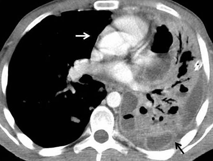
Also, CT revealed a loculated left pleural effusion with thickened and enhanced visceral and parietal pleura (the split pleura sign), suggestive of empyema (Fig. 3). There was no lymphadenopathy.

Chest CT showing left loculated pleural effusion with thickening and enhancement of parietal and visceral pleura (black arrow). In addition, ipsilateral mediastinal shift is produced (white arrow).
Fig. 3: Chest CT showing left loculated pleural effusion with thickening and enhancement of parietal and visceral pleura (black arrow). In addition, ipsilateral mediastinal shift is produced (white arrow).

Differential Diagnosis List

  • Hydropneumothorax as the initial manifestation of postprimary tuberculosis.
  • Chronic obstructive pulmonary disease (emphysema, cystic fibrosis…)
  • Lung cancer
  • Other infection (coccidioidomycosis, aspergillosis, histoplasmosis…)
  • Pneumocystis jiroveci (in HIV-related disease)

SIMILAR CASE: Hydropneumopericardium After Intubation