ECG Interpretation
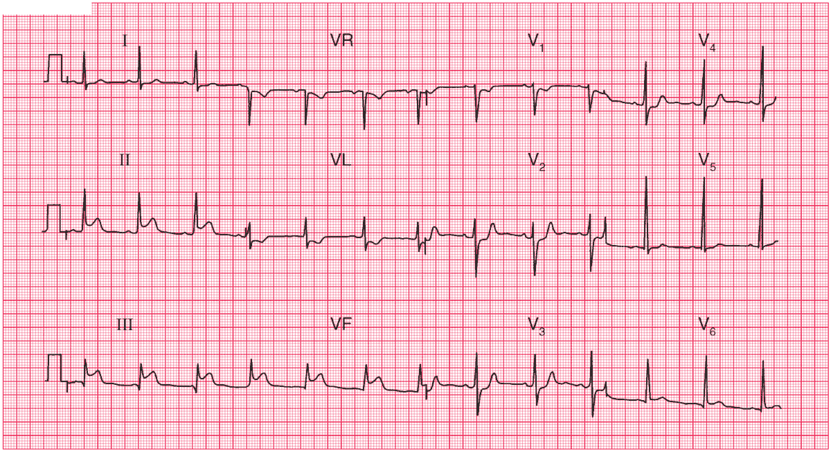
- Sinus rhythm, rate 72/min
- Normal axis
- Small Q waves in lead III
- Elevated ST segments in leads II, III, aVF, with upright T waves
- Reciprocal ST segment depression in Leads I and aVL
- ST segment depression in leads V1–V4
- T wave inversion in lead aVL
Clinical Interpretation
The ST elevation in inferior leads (II, III, aVF) with reciprocal ST depression in lateral leads (I and aVL) tells us this man has Inferior MI. The ST depression in leads V1-4 shows us there is also Posterior MI. Because the ST Segment in V1 is not isoelectric or elevated, Right Ventricular infarction is unlikely.
What to do next ?
In the absence of contraindications (i.e. risk of bleeding from any important site), the patient should be given aspirin and nitroglycerin and then percutaneous coronary intervention (PCI) or a thrombolytic agent.


