ECG Interpretation
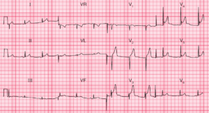
- Sinus rhythm, rate 79/min
- Right axis deviation
- Normal QRS complexes, except for an RSR1 pattern in lead V1 and deep S waves in lead V6
- Inverted T waves in leads V1–V4 (right ventricular strain pattern)

Clinical Interpretation
The right axis deviation, the deep S waves in lead V6 (‘clockwise rotation’) and the inverted T waves in the right chest leads (V1 to V4) are all characteristic of right ventricular strain in this case due to pulmonary embolism.
The pulmonary angiogram showed clots within the main pulmonary arteries.
What to do ?
In the context of a delivery 3 months previously, this ECG pattern of right ventricular strain almost certainly indicates multiple pulmonary emboli causing pulmonary hypertension. The pulmonary angiogram confirms this diagnosis. Anticoagulants, and possibly thrombolysis, are needed urgently.