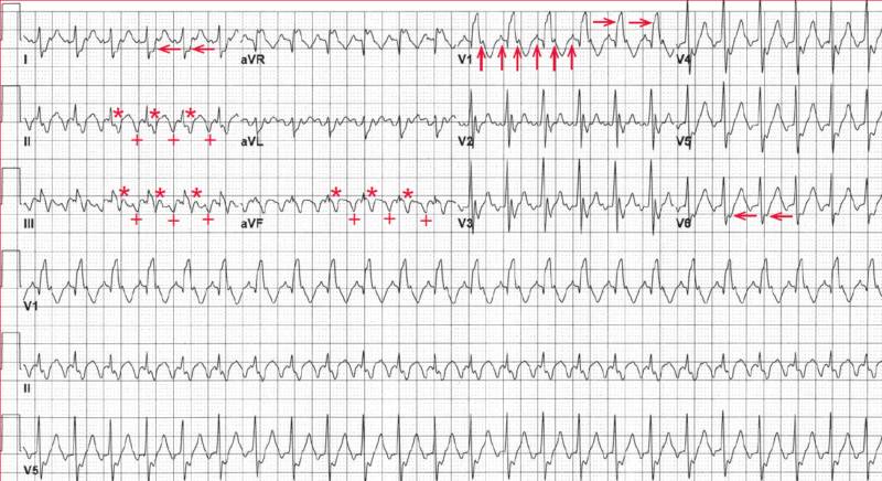
The rhythm is regular at a rate of 150 bpm. The QRS complex is widened (0.14 sec), and there is an RSR′ morphology in lead V1 (→) and a broad S wave in leads I and V5-V6 (←); this is right bundle branch block (RBBB).
The axis is normal (positive QRS complex in leads I and aVF, ie, between 0° and +90°). The QT/QTc intervals are normal (280/440 msec and 220/350 msec when corrected for the prolonged QRS duration).
A negative atrial waveform can be seen before each QRS complex in leads II, III, and aVF (+). An identical atrial waveform can be seen at the end of the QRS complex in these leads (*). In addition, a distinct atrial waveform before and after each QRS complex can be seen in lead V1 (↑).
The interval between these waveforms is constant, and the atrial rate is 300 bpm. There is no isoelectric baseline between these waveforms; rather the baseline is constantly undulating (saw-tooth pattern). Hence this is atrial flutter with 2:1 AV conduction.
- READ MORE: Atrial Flutter: ECG Interpretation [With Examples]
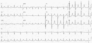
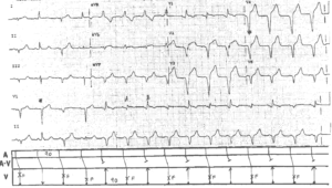
- Similar Cases:


