This post is an answer to the ECG Case 225
- Rate: ~90 bpm
- Rhythm:
- Regular
- Sinus Rhythm
- Last complex PVC
- Axis: LAD
- Intervals:
- PR – Prolonged (220ms)
- QRS – Normal (100ms)
- QT – 320ms (QTc Bazette 380-400 ms)
- Segments:
- ST elevation in leads III, V1-5
- ST depression (minor) in lead I
- Additional:
- Q wave in V1-2
- QS Wave in V3-4
- T inversion in leads aVR, aVL
- Voltage criteria LVH – aVR ~1.2mV
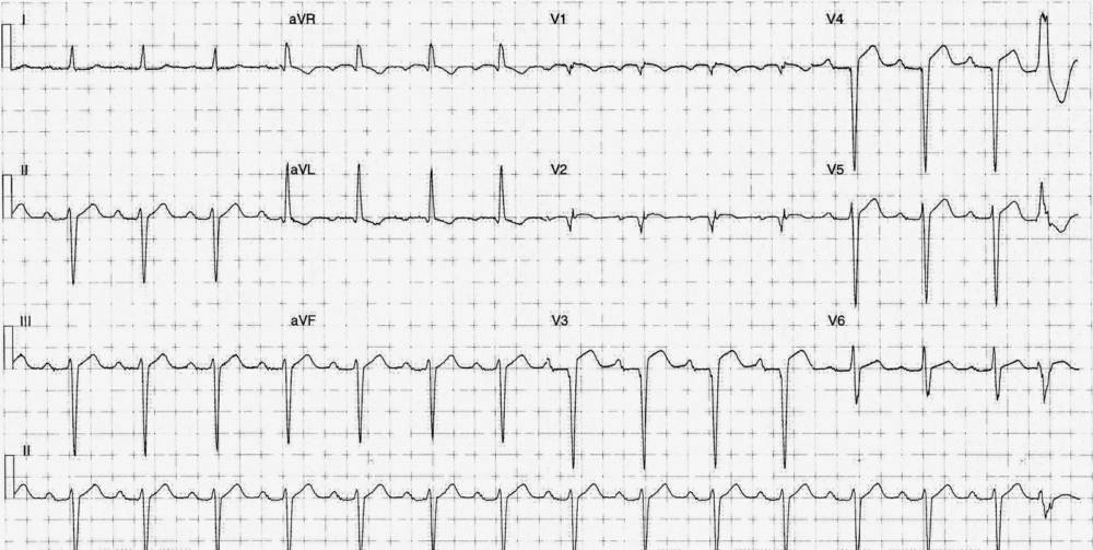
Interpretation:
ECG features stongly suggest an old anteroseptal MI, with Q / QS waves in precordial leads, a lack of significant ST depression, and a pain-free patient with a known previous ischaemic insult. This patient was admitted and investigated for dyspnea further with the following investigations:
Myocardial Perfusion Scan
- Large anteroseptal and apical infarction
- Small area of reversible ischaemia in the mid-to-basal anterior wall
Echocardiography
- Severely impaired systolic function
- Large anteroapical aneurysm
- No LV thrombus
- LV ejection fraction 27%
READ MORE: Know the Differential for ST Segment Elevation: It’s More Than Just ACS
SIMILAR CASES:
![Read more about the article Hypokalemia ECG Changes [With Examples]](https://manualofmedicine.com/wp-content/uploads/2021/04/Hyperkalemia-and-Hypokalemia-ECG-Changes-2-300x127.jpg)