A 22-year-old man presented to the hospital after a motor vehicle collision in which his head struck the windshield of a bus. Initial observations and the physical and neurologic examinations were normal, but the injury warranted computed tomographic (CT) scanning.
A CT scan of the brain (Panel A) showed an incidental finding of grossly dilated occipital and temporal horns of the right lateral ventricle.
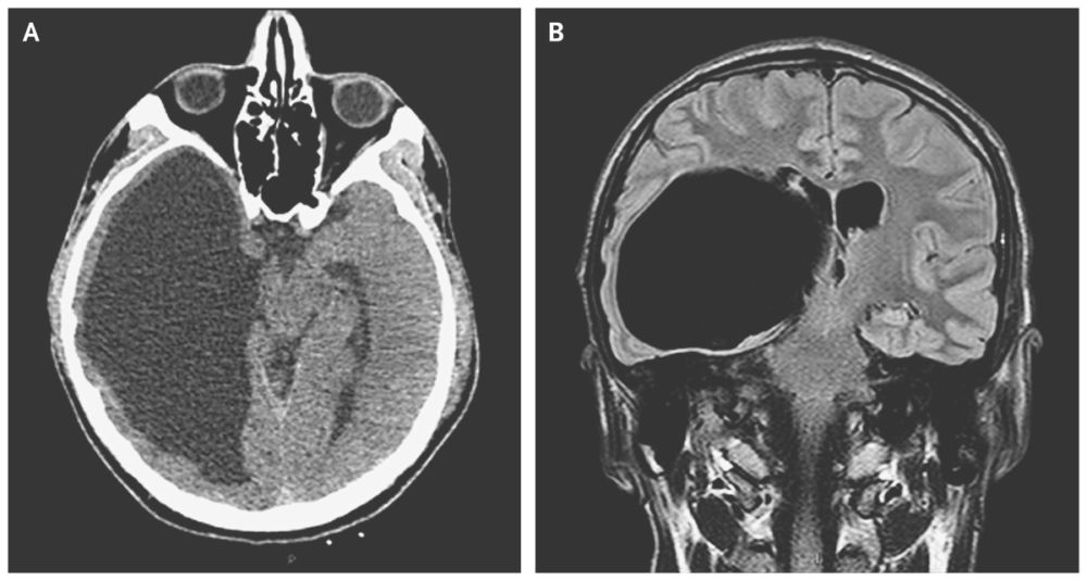
Magnetic resonance imaging (Panel B) confirmed the presence of a large cystic lesion extending from the temporal and posterior horns of the right lateral ventricle (axial size, 11 cm by 7 cm), with a mild midline shift of the third ventricle, and compression of the midbrain and brain stem with thinning of the temporal and occipital cortexes. A large, asymptomatic, intraventricular arachnoid cyst was diagnosed.
Arachnoid cysts — collections of cerebrospinal fluid within the layers of the arachnoid membrane — occur infrequently. Most are congenital, but they can also be acquired after trauma or infection through the entrapment of cerebrospinal fluid within arachnoid adhesions.
Neurosurgery was recommended in this case, but the patient discharged himself from the hospital and there was no further follow-up.