
This article is an answer to the Case – A 72-year-old Woman with Abdominal Pain, Nausea and Vomiting
On physical examination, there were no abdominal scars or umbilical, inguinal, or femoral hernias. Laboratory tests revealed a normal white-cell count, and an abdominal radiologic examination was suggestive of a complete small-bowel obstruction (Panel A).
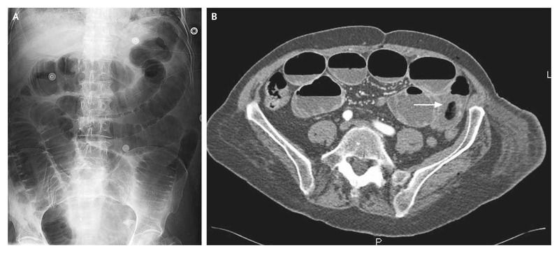
Computed tomography showed small-bowel obstruction by an intraluminal mass (Panel B, arrow). This mass had a hyperdense periphery and an aerated core.
During laparotomy, an enterotomy was performed and a plastic ball was found within the lumen. The ball was 4 cm in diameter and had a hard plastic layer and a soft core.
Additional questioning did not reveal whether the ingestion had been voluntary or accidental or when it might have occurred. The patient had an uneventful recovery.