Table of Contents
Annually, almost 6 million patients present to the emergency department with a chief complaint of chest pain. Thankfully, the majority will not have an acute coronary syndrome (ACS) as the etiology of their symptoms. There are numerous etiologies of chest pain that range from benign to life threatening.
The emergency provider (EP) should broaden the differential of acute chest pain beyond simply ACS, in order to promptly recognize and treat additional life-threatening etiologies of chest pain.
Life-threatening causes of acute chest pain include:
- ACS ( Acute coronary syndrome )
- Aortic dissection
- Pulmonary embolism (PE)
- Tension pneumothorax
- Cardiac tamponade
- Esophageal rupture.
Delayed diagnosis of these etiologies is associated with significant increases in patient morbidity and mortality.
Thoracic Aortic Dissection (TAD)
The incidence of thoracic aortic dissection (TAD) is ~3 cases per 100,000 people per year. The incidence of TAD peaks at age 70 and is more common in males. Patients commonly have a history of hypertension. Patients with TAD who are younger than 40 years of age commonly have a history of:
- Connective tissue disease (i.e., Marfan syndrome, Ehlers-Danlos syndrome)
- Chronically abuse cocaine
- History of a bicuspid aortic valve
Additional risk factors for TAD include:
- Pregnancy, especially in the third trimester
- Prior history of aortic surgery
- Aortic valve disease
- Family history of aortic valve disease.
Presentation of TAD
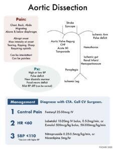
The most common presenting symptom is the abrupt onset of severe pain. In contrast to the textbook descriptions of “tearing” or “ripping” pain, patients with TAD more commonly report the abrupt onset of sharp pain that quickly reaches maximal intensity. TAD should also be considered in any patient in whom symptoms cross the diaphragm (i.e., chest and abdominal pain). Acute onset of thoracic back pain can be reported in patients with a dissection of the descending aorta.
Physical Examination
Classic physical examination findings in the patient with TAD include:
- Blood pressure differential between the extremities
- Extremity pulse deficit
- Aortic insufficiency murmur
- Focal neurologic finding.
Importantly, these findings are variable. In fact, a systolic blood pressure differential has been found to be both poorly specific and sensitive for TAD. A normal physical examination should not exclude the diagnosis of TAD when the history is strongly suggestive of this etiology.
Diagnosis of TAD
Chest x-ray (CXR) abnormalities seen in patients with TAD include a widened mediastinum and an abnormal aortic contour. These classic findings, however, are infrequently found in the majority of cases. Computed tomography (CT) angiography is commonly used to confirm, or exclude, the diagnosis of TAD. In recent years, there have been numerous studies that have attempted to validate the use of D-dimer in the evaluation of patients with TAD. At present, a negative D-dimer is insufficient to exclude TAD in low-risk patients.
Pulmonary Embolism (PE)
Read more about Pulmonary Embolism (PE) here.
The estimated incidence of PE continues to rise. Risk factors for PE are well documented and include a history of hypercoagulability, recent surgery or prolonged immobilization, connective tissue disease, and exogenous estrogen use.
Patients with PE commonly report dyspnea, with or without exertion, and acute chest pain. The chest pain of PE is typically described as pleuritic in character. Other historical features that may guide the EP are the presence of lower extremity or calf pain, unilateral lower extremity swelling, cough, or hemoptysis.
Similar to TAD, physical examination findings are often absent in the patient with PE. Vital sign abnormalities may raise the clinician’s suspicion, but are not always present at the time of ED evaluation. Patients with PE can have tachycardia, tachypnea, or hypoxia. With a massive PE, the patient may present with hypotension and signs of shock.
Fever, when present, is typically low grade and can mislead the EP toward a diagnosis of pneumonia. Unilateral lower extremity swelling can suggest the diagnosis of DVT and raise suspicion for PE.
The diagnostic evaluation for PE requires an understanding of current clinical decision rules. The well-versed EP should be able to exclude PE in low-risk patients with a combination of clinical gestalt and the Pulmonary Embolism Rule-out Criteria.
Beyond these low-risk patients, EPs must understand and correctly apply the Well’s criteria or Revised Geneva Score to calculate pretest probability and guide further evaluation with either a Ddimer test or CT angiography of the chest.
Tension Pneumothorax
The incidence of tension pneumothorax varies widely and is dependent on the population studied. The EP should have a high index of suspicion for tension pneumothorax in patients with a history of trauma or recent instrumentation of the thorax, neck, or upper extremity regions.
Tension pneumothorax almost always presents with the combination of acute chest pain and respiratory distress.
Clinical features include unilateral or absent breath sounds, hypotension, jugular venous distension, and tracheal deviation.
Tension pneumothorax is a clinical diagnosis that requires rapid intervention to prevent cardiac arrest and death. Bedside ultrasound can be used to quickly confirm the diagnosis in patients with an equivocal exam.
Cardiac Tamponade
In cardiac tamponade, a pericardial effusion increases pericardial pressure, decreases right ventricular filling, and decreases cardiac output.
Etiologies of pericardial effusion include:
- Malignancy
- Trauma
- Infectious diseases
- Pericarditis
- Uremia
- Acute myocardial infarction.
Patients with a pericardial effusion with development of tamponade often report chest pain, dyspnea, and fatigue.
Diminished breath sounds or a pericardial friction rub are heard in only one-third of patients.
The classic triad of hypotension, muffled heart sounds, and jugular venous distension is a late finding in patients with tamponade.
The electrocardiogram in patients with a pericardial effusion can demonstrate:
- Low voltage
- Tachycardia
- Electrical alternans
Similar to tension pneumothorax, bedside ultrasound can be used to quickly confirm the diagnosis. The presence of an effusion with diastolic right ventricular collapse should lead to emergent pericardiocentesis
Esophageal Rupture
Esophageal rupture is a rare diagnosis. Common precipitants of esophageal rupture include:
- Iatrogenic (i.e., esophagogastroduodenoscopy)
- Severe emesis
- Trauma
- Caustic ingestion
- Esophageal foreign body.
Chest pain is most often retrosternal, is severe, and frequently radiates to the back, neck, shoulders, or abdomen. Additional historical features may include dysphagia, dyspnea, and emesis.
Patients with esophageal rupture can present in shock with tachycardia, hypotension, and signs of poor perfusion.
Physical exam findings can include subcutaneous emphysema in the cervical and clavicular region. Patients with an intra-abdominal rupture may present with signs of a surgical abdomen.
Although a CXR can demonstrate a pneumothorax, pneumomediastinum, or pleural effusion, the diagnosis of esophageal rupture is confirmed with CT of the chest. CT findings of esophageal rupture can include periaortic or periesophageal air, pleural effusions, or soft tissue stranding.
Key Points
- Patients with TAD more commonly report the abrupt onset of sharp pain that quickly reaches maximal intensity.
- Patients with PE may not present with tachycardia, tachypnea, or hypoxia.
- Tension pneumothorax remains a clinical diagnosis.
- The classic triad of hypotension, muffled heart sounds, and jugular venous distension is a late finding in patients with cardiac tamponade.
- The most common etiology of esophageal rupture is iatrogenic.


