This article is an answer to the ECG Case 180
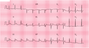
The ECG shows the rhythm is regular at a rate of 160 bpm. The QRS complex duration is normal (0.08 sec). The QRS complex morphology is normal, although there is an R′ in lead V1 (←), representing a minor right ventricular conduction delay. This QRS complex morphology in V1 has been termed a crista pattern, as the last portion of the right ventricle to depolarize is the crista supraventricularis.

The axis is about 0° (positive QRS in lead I and biphasic QRS complex in lead aVF). The QT/QTc intervals are normal (270/440 msec). There are no obvious atrial waveforms seen in any lead. However, there are uniform and regular positive waveforms seen before each QRS complex in leads II, aVF, and V1 (+). In leads III and aVF these waveforms appear to be negative–positive. This is a long-RP (short PR) tachycardia (RP interval = 0.24 sec, PR interval = 0.12 sec).
Etiologies for a long-RP tachycardia include sinus tachycardia, atrial tachycardia, ectopic junctional tachycardia, atypical atrioventricular nodal reentrant tachycardia, atrioventricular reentrant tachycardia or atrial flutter with 2:1 AV block.
The P waves seen are negative positive in leads III and aVF, and hence the rhythm is not sinus. With careful inspection of lead aVF, it can be seen that there is a second waveform of the same axis and morphology at the end of the QRS complex (^). The interval between the waveform before and after the QRS complexes in lead aVF is regular at a rate of 320 bpm (┌┐). These findings are consistent with atrial flutter with 2:1 AV conduction.

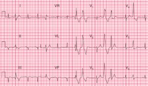
This second ECG confirms the diagnosis. In leads II, III, and aVF these waveforms are negative positive and have a “sawtooth” pattern. In lead V1 the waveforms are positive–negative (^). Hence this is typical atrial flutter with variable block (3:1 and 4:1), ultimately becoming stable with 2:1 AV block.
READ MORE: Atrial Flutter – ECG Interpretation [With Examples]
SIMILAR CASES: