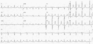
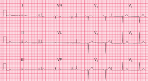
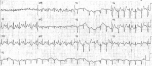
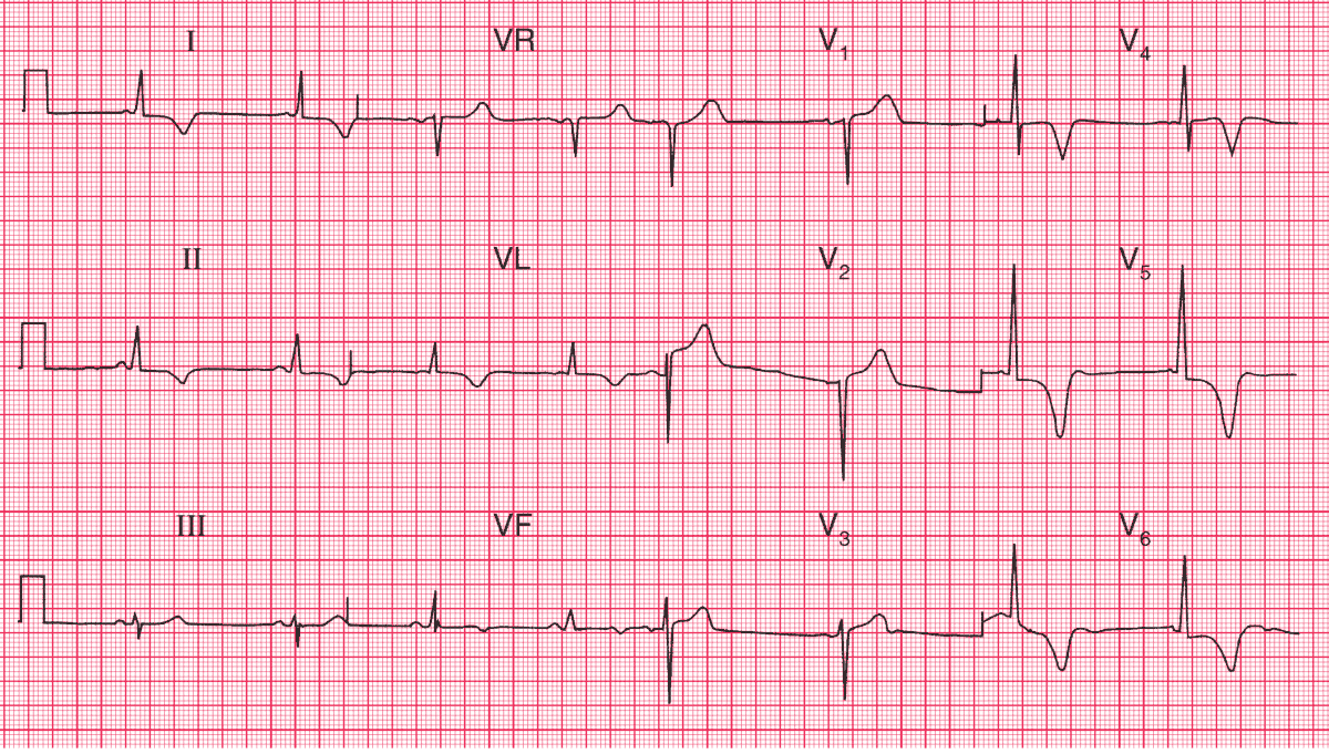
ECG Interpretation
- Sinus rhythm
- Normal axis
- Normal QRS complexes
- Marked T wave inversion in leads I, II, VL, V4–V6
Clinical Interpretation
Anterolateral T wave inversion as gross as this may be due to a non-ST segment elevation myocardial infarction, or even to left ventricular hypertrophy.
However, there are no other features of left ventricular hypertrophy on this trace, which is fairly characteristic of hypertrophic cardiomyopathy. Myocardial infarction is uncommon in people of this age.
What to do ?
Physical signs of hypertrophic cardiomyopathy include a ‘jerky pulse’; an aortic flow murmur which is characteristically louder after the pause that follows an extrasystole; and mitral regurgitation.
Read More about Heart Murmurs – Differential Diagnosis, Examination and Investigations
Hypertrophic cardiomyopathy is best diagnosed by echocardiography, which will show asymmetric septal hypertrophy, systolic anterior movement of the mitral valve apparatus, and sometimes early closure of the aortic valve.
This patient’s echocardiogram showed all these features, confirming the diagnosis of hypertrophic cardiomyopathy.
- READ MORE:
- Similar Cases: